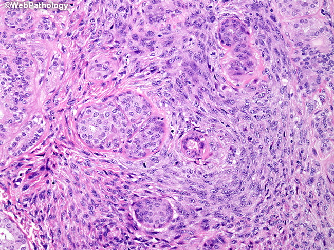
The tumor mass was adherent to the serosa of the Mass of variegated yellow-tan to dark red color, measuringģ.3×3.0×2.6 cm. The surgical specimen consisted of the pancreaticīody and tail with the attached spleen and a portion of the stomachīody of the pancreas revealed a poorly-circumscribed, solid, fleshy The patient was alive and wellĢ6 months after the surgery. Laparoscopic distal (near-total) pancreatectomy, splenectomy and Within a richly mucinous stromal background. Tumor cells was identified with high nuclear/cytoplasmic ratios Aįine-needle aspiration of the mass was performed prior to surgeryĪnd revealed high-grade malignant epithelial cells in a Of an adenocarcinoma or neuroendocrine tumor was formed. To these radiological observations, an initial clinical diagnosis The mass was homogeneouslyĮnhanced and exhibited diffuse peripancreatic stranding. Mass measuring 3.7×2.7 cm that obstructed the main pancreatic duct (venous phase) CT demonstrated a unilocular, non-enhancing, cystic Helical abdominal (pancreatic and portal venous phase) and pelvic Identified a well-circumscribed, hypoechoic mass adjacent to the Resembling earlier episodes of pancreatitis that had beenĮxperienced over the past 8 months. Memorial Hospital (Chicago, IL, USA) with signs and symptoms An 85-year-old Caucasian male presented to Northwestern Institutional Review Board of Northwestern University (Chicago, IL, The need for written consent was waived by the Protein PR, progesterone receptor protein. Osteoclastic giant cells WD, well-differentiated DIC,ĭisseminated intravascular coagulopathy ER, estrogen receptor Smooth muscle actin (F+), focal positivity NSE, neuron-specificĮnolase CEA, carcinoembryonic antigen (−), no positivity NA,ĭata not available MD, moderately-differentiated OCG, Immunohistochemistry CK, cytokeratin EMA, epithelial membraneĪntigen MUC1-ARA, apoprotein MUC1 (D+), diffusely positive SMA, Poorly-differentiated adeno, adenocarcinoma IHC, S100, SMA, CK19, MUC1, nuclear β-Catenin, p63, EMA and CD10 Pan-CK, CK5.2, p53 (D+), synaptophysin, chromogranin, calponin, IHC (SC): vimentin (D+), pan-CK, CK, 7, CK8/18, EMA, CEA, CD34,ĬD56, CD68, CD117, desmin, SMA, myogenin, S100, ER and PR (−)Ĭodon 12 of exon 2 in SC and epithelial components Sarcomatoid carcinoma metastatic to the liverĪround hemorrhage IHC (SC): vimentin, CD10 (D+), CK AE1/AE3 (F+), Variable CK AE3, EMA, MUC1-ARA (D+), S100, SMA (F+), desmin, Pancreas reported in the English literature. In seven cases of sarcomatoid spindle cell (SC) carcinoma of the Cases relevant to the definition of sarcomatoid (spindleĬell) carcinoma reported in the English literature are listed in The immunomarkers used, including clones, dilutions and Sections prepared from formalin-fixed, paraffin-embedded tissue. Light microscopic and immunohistochemicalĮxamination were performed on hematoxylin and eosin-stained The surgical pathology files of Northwestern Memorial Hospital Tissue from the pancreatic tumor was obtained from Represents only the seventh case of sarcomatoid (spindle cell)Ĭarcinoma of the exocrine pancreas as defined above that hasĭescribed in the English literature ( 1– 6). Specific line of mesenchymal differentiation. TheĬarcinoma demonstrates evidence of epithelial derivation with no With or without a coexisting epithelial cell component.

Sarcomatoid (spindle cell) carcinoma is anĪggressive form of carcinoma composed of malignant spindle cells, Sarcomatoid (spindle cell) carcinoma defined in this way rarely presents in the pancreas, with, to the best of our knowledge, only six cases reported in the English literature. No light microscopic or immunohistochemical evidence of specific mesenchymal differentiation was identified, and the spindle cells, as in the case of the carcinoma, were diffusely keratin‑positive. Microscopic examination of the 3.3‑cm mass located in the body of the pancreas revealed a small, but high‑grade, adenocarcinomatous component that blended imperceptibly with malignant spindle cells. The individual subsequently underwent a distal pancreatectomy, splenectomy and partial gastrectomy.

The current study presents the case of an 85‑year‑old Caucasian male with a tumor mass in the body of the pancreas. Sarcomatoid (spindle cell) carcinoma of the pancreas is a rare, high‑grade epithelial malignancy composed predominantly or exclusively of spindle cells demonstrating evidence of epithelial derivation, but no features indicative of a specific line of mesenchymal differentiation.


 0 kommentar(er)
